|
|
|
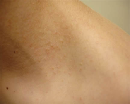
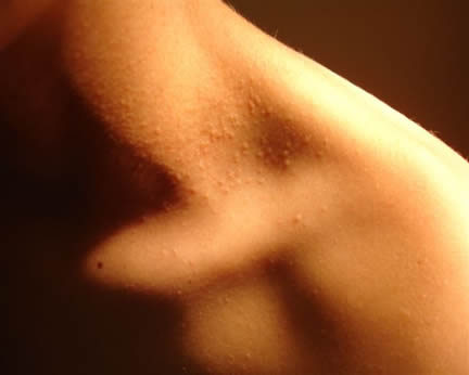
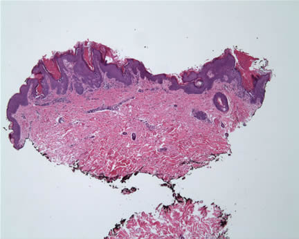
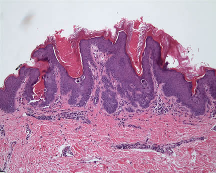
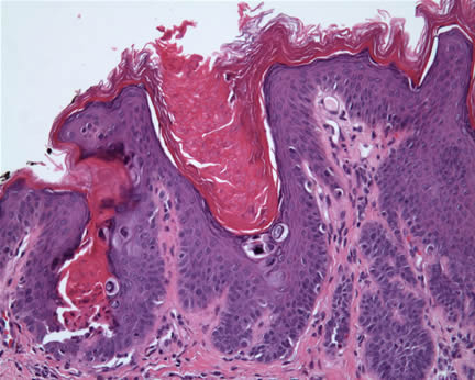
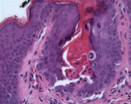
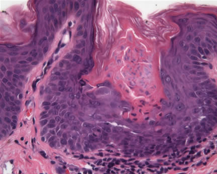
Case Study Diagnosis- Comment: Darier's disease is a genodermatoses. It classically involves the seborrheic areas of the face, scalp, and trunk. Patients occasionally present with nail dystrophy and other abnormalities. The skin biopsy shows characteristic findings of an acantholytic dermatosis with corp ronds and corp grains. In most cases, the clinical and histopathologic findings are straightforward. In this case, both the clinical and histopathologic findings were a bit unusual. The clinical lesions lacked the typical appearance of brown, greasy warty papules but were instead flesh colored smooth papules. They were also localized to one side of the neck with no lesions on the hands or nails. Linear or segmental clinical variants have been reported and these cases may be associated with genetic mosaicism of ATP2A2. Mutations in this latter gene have been implicated in the pathogenesis. The gene normally encodes for the calcium pump located in the sarcoplasmic/endoplasmic reticulum Ca2+-ATP isoform 2 protein (SERCA2). Of interest in confirming the diagnosis, oral corticosteroids may worsen the disease. It is interesting that this patient's lesions worsened with topical steroid treatment. This patient was presented to the University of California Irvine Dermatology Grand Rounds where a consensus diagnosis of Darier's disease was reached. Case and Clinical Photos Submitted by: References: |
First Posted August 31, 2006
Send Emails to
Webmaster at DermpathMD
Read the Medical Disclaimer