|
|
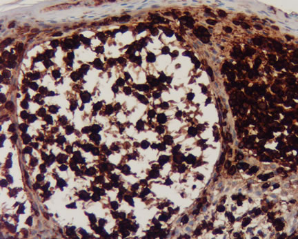
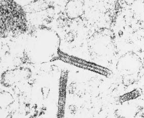
Figure 4-Langerhans cells are strongly positive for CD1a. Note the benign non-staining squamous epithelial cells. |
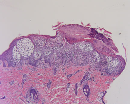
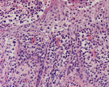
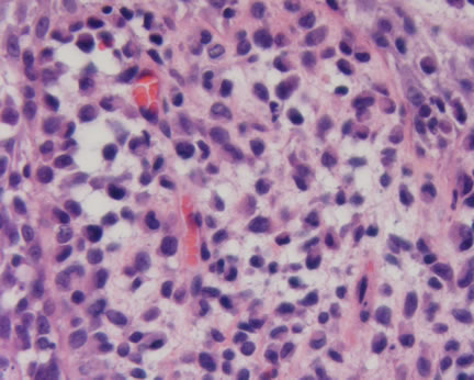
Case Study This is a 3 1/2 month old boy with a one month history of a scaly papular rash on the scalp and trunk. Diagnosis: Langerhans Cell Histiocytosis This is an uncommon hematopoietic neoplasm of the skin. For many years, these diseases were known by such terms as Histiocytosis X, Hand-Schuller-Christian disease, Eosinophilic granuloma, and Letterer-Siwe disease. Currently, all of these previous designations are considered clinical variants, best categorized under the appellation cited above. The unifying histopathologic feature is the Langerhans cell, a cell normally involved in antigen processing within the skin. These cells have a characteristic immunohistochemical profile for CD1a (Figure 4) and for S100. If one can avail of an electron microscope, a diligent search will reveal the Birbeck granule (Figure 5), pathognomonic for a Langerhans cell. The clinical presentation of this patient is rather classic but as we all know, sometimes tumors do not read the books. The recent dermatology literature is replete with unusual clinical presentations, including a blueberry muffin baby (see reference below). Clinical photograph and information submitted by Elizabeth Lener, M.D. References: Braier J, Chantada G, Rosso D, Bernaldez P, Amaral D, Latella A, Balancini B, Masautis A, Goldberg J. Langerhans cell histiocytosis: retrospective evaluation of 123 patients at a single institution. Pediatr Hematol Oncol 1999 Sep-Oct;16(5):377-85. Howarth DM, Gilchrist GS, Mullan BP, Wiseman GA, Edmonson JH, Schomberg PJ. Langerhans cell histiocytosis: diagnosis, natural history, management, and outcome. Cancer 1999 May 15;85(10):2278-90.
Archived Case Studies |
Last Updated July 29, 2005
Send Emails to
Webmaster at DermpathMD
Read the Medical Disclaimer