
|
|
|
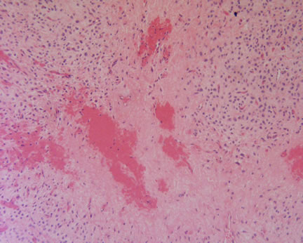
Case Study This is an occipital scalp lesion arising in a 49 year old man. There is a vague history of trauma at this site a few months earlier. Diagnosis: Ossifying Fibromyxoid Tumor This is a rare soft tissue neoplasm usually characterized by a small, painless, well-circumscribed mass in the subcutis or muscle. The most frequently reported locations include the upper and lower extremities, trunk, and head and neck region. Rarely cutaneous cases have been reported. The tumor has a characteristic combination of small, round cells arranged in a cord- or nest like pattern within a myxoid matrix. Transitions with areas of hyaline fibrosis and osteoid formation are frequent. The cells are often positive for S-100 protein, a feature found in this case. The prognosis is guarded and the tumor is currently classified as a sarcoma of intermediate malignant potential. In the initial series, no metastases or deaths were recorded although local recurrence occurred in about 27% of cases. Subsequent papers have documented malignant cases and death from the disease. The origin of these tumor cells is still uncertain with most studies suggesting a cartilaginous or neural origin. References: J Am Acad Dermatol. 2005 Apr;52(4):644-7 Archived Case Studies |
Last Updated April 25, 2005
Send Emails to
Webmaster at DermpathMD
Read the Medical Disclaimer
 Ossifying Fibromyxoid Tumor