| Malignant Melanoma, Reporting Template
The information in melanoma pathology report should closely reflect the clinical prognosis. Several items should be clearly addressed. These include the depth of invasion, the level of invasion, the presence or absence of ulceration and the margin.
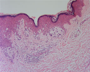
Depth of invasion (Breslow’s thickness)
This is the single most important factor in predicting survival for melanoma patient. It is measured from the top of the granular layer to the deepest tumor extension.
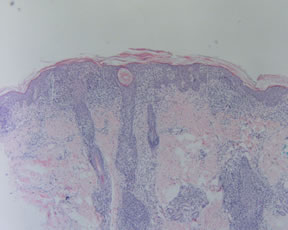
Level of invasion
This has descriptive value. The different levels appear to reflect the sequential acquisition of new properties by evolving tumors. In level I tumor (melanoma in-situ), the melanoma cells are confined to the epidermis and its appendages Tumor cells extent into the papillary dermis in level II tumor and throughout the papillary dermis in the level III tumor. Once the melanoma cells invade reticular dermis or subcutaneous fat, it became a level IV or V tumor.
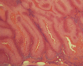
Ulceration
Recently, ulceration is used in the T classification in major revisions of the melanoma TNM and stage grouping criteria by the Melanoma Staging Committee of the AJCC.
Margin
Generally speaking, a margin of 0.5-cm is adequate for malignant melanoma in-situ. A 1.0-cm margin is sufficient for low risk melanoma up to 1mm in thickness and 2-cm margin for tumors with 1-4 mm thickness. For lesions greater than 4-mm, 2- to 3-cm margin is generally recommended.
In level II melanoma, the presence or absence of the vertical growth phase (VGP should be mentioned, since they carry different prognostic results. The important and useful criterion of VGH is the presence of dermal mitosis or small expansile nodule that is conspicuously larger than any nest within epidermis.
Other Histological Features
These features,
if present, should also be stated in the report. These include regression, tumor-infiltrating lymphocytes, mitotic rate, vascular or lymphatic invasion, precursor lesion (dysplastic or congenital nevi) and satellites.
Submitted by Yong Tao, M.D., PhD.
References:
Balch CM, et al, A new American Joint Committee on Cancer staging system for cutaneous melanoma. Cancer. 2000 Mar 15;88(6):1484-91.
Balch CM, Buzaid AC, Soong SJ, et al: Final version of the American Joint Committee on Cancer staging system for cutaneous melanoma. J Clin Oncol 19:3635-3648, 2001
Balch CM, Soong SJ, Gershenwald JE, et al: Prognostic factors analysis of 17,600 melanoma patients: Validation of the American Joint Committee on Cancer melanoma staging system. J Clin Oncol 19:3622-3634, 2001 |