
|
|
Image 1-In some areas, the tumor is composed of cellular nodules admixed with areas of hyalinization. In other foci, prominent bone formation is noted.
|
Case Study
This is an occipital scalp lesion arising in a 49 year old man. There is a vague history of trauma at this site a few months earlier. |
Last Updated April 25, 2005
Send Emails to
Webmaster at DermpathMD
Read the Medical Disclaimer
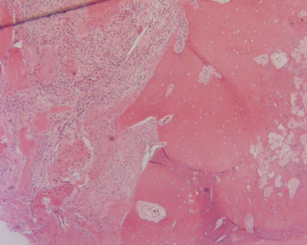
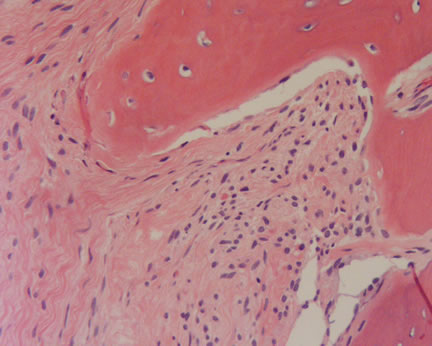
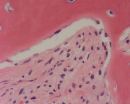
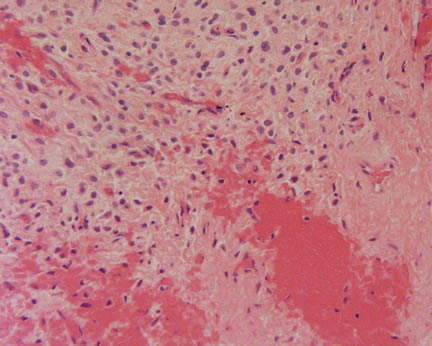 Image 4-Another focus shows the spindle cells merging with areas of hyalinization.
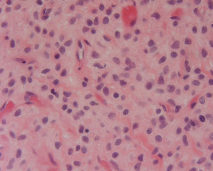 Image 5-Highest magnfication reveals cytologic blandness of the spindle cells.